our research
our research focuses on the interplay between biomedical imaging, biomechanics, and device design. we work in the vascular, respiratory, and hepatic space and have a strong interest in women’s breast and reproductive tissue health. by working closely with clinicians, this work originates in ex vivo research with the goal for its eventual clinical translation.
-
women's health
harnessing our expertise in imaging, biomechanics, and device design we are looking towards understanding reproductive tissues and breast tissues in order to innovate within women's healthcare.
-
device design
with improved visualisations into tissue-device interactions from non-invasive imaging, we look to use this insight to design better devices as well as monitor their integration.
-
imaging
with a focus quantitative magnetic resonance imaging and ultrasound methods, we aim to use clinically relevant imaging to characterise the microstructure and microenvironment of tissue
-
biomechanics
coupled with our imaging focus on microstructural components, our group looks to understand the mechanical implications of component content and alignment, as informed by imaging.
explore our vacancies
*
explore our vacancies *

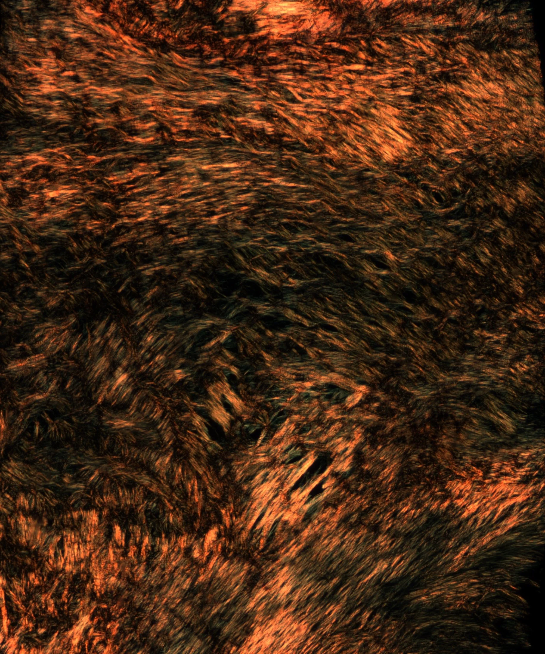
diffusion tensor imaging
diffusion tensor imaging is a magnetic resonance imaging technique which uses the diffusion of water molecules to map a tissue’s underlying microstructure. with extensive work done in arterial tissue, we have seen the sensitivity of this method to cell and elastin content. our more recent work looks to investigate this method in liver tissue, as well as breast and reproductive tissues.
Right: diffusion tensor imaging derived tractography of excised human carotid artery (https://doi.org/10.1161/ATVBAHA.122.318112)
ongoing funding, projects + collaborators
-
![]()
NOVAP: novel device for ventilator associated pneumonia prevention
Funded by an Enterprise Ireland Commercialisation Fund
-
![]()
Irish Research Council Employment based PhD
Co-funded by Stryker Neurovascular
-
![]()
Trinity St. James’s Cancer Institute Cancer Research Stimulus Awards
The TSJCI CREST Awards are supported by a philanthropic gift from The Dr Margaret Sau Sheung Ip and Dr Jonathan Chiu Fund.
-
![]()
Trinity College Dublin
TCD Trust Grants
-
![]()
Trinity Research Doctoral Awards
PI-led TRDA (‘25-’26)
-
![]()
earlier.org - Friends for an Earlier Breast Cancer Test
Unmasking breast collagen with MRI